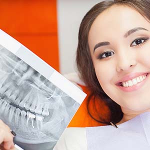
Periapical X Rays
Periapical X-rays capture an image of the whole tooth from the crown above the gumline to the root end. Used to highlight only one or two teeth at a time, they provide a top to bottom view that helps diagnose painful conditions or changes in teeth resulting from periodontic treatments.
Taking a periapical X-ray requires placing a small strip of film in a plastic holder into the mouth. Attached to the holder is a frame with a ring that mirrors the position of the film, helping the dental assistant align the X-ray machine opposite the tooth. Patients bite down on the holder to keep the film steady while taking the X-ray.
Periapical X-rays are especially useful for discovering and determining the best course of treatment for difficult oral health problems like impacted teeth, cysts, abscesses, and even tumors.
Periapical X-rays can be used to target individual areas of the mouth, as well as the full mouth, and are usually taken in a longer series. The benefits of having two methods of radiography available ultimately amount to more effective diagnostic capability.
If you are experiencing a toothache or if there is a suspicious-looking area on one of your teeth, we would take periapical X-rays to get a better idea of what is going on. With these x-rays, dentists are also able to see the root structure of the tooth as well.
Dentists also recommend having periapical x-rays taken once a year when your 4 bitewing x-rays are taken. In this situation, dentists would take them to evaluate the health of your front teeth, which are not included in the bitewing series.
#periapical x rays
.
